Tracheostomy
There are two airways, not one. Unlike a laryngectomy, the mouth may still work - so oxygen goes on BOTH the face and the stoma.
Also called: trache, trachy, neck breather, surgical tracheostomy, percutaneous tracheostomy, green algorithm patient

What it is
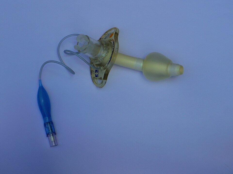
A tracheostomy is a tube through an opening in the front of the neck into the trachea. Crucially, and unlike a laryngectomy, the upper airway is usually still intact: the mouth and nose remain connected to the lungs, so oxygen and ventilation at the face may still work. That single fact is why the NTSP publishes a green algorithm for tracheostomy and a separate red one for laryngectomy.
But 'usually' is not 'always'. When the tracheostomy was formed for an obstructing head-and-neck tumour, laryngotracheal stenosis, or after a partial laryngeal or tracheal resection, the upper airway may be impassable and attempts at the face will fail. If oxygen and intubation at the face do not work quickly, move to the stoma without delay - and ask why the tracheostomy was formed.
The tube may be surgical or percutaneous, cuffed or uncuffed, with or without an inner cannula, and may carry a speaking valve or cap. How and when it was formed changes how safely it can be replaced - see below.
How to recognise it
- A tube in the front of the neck, held by tapes or a dedicated holder, often with a visible inner cannula and sometimes a speaking valve or cap on the end.
- A green NTSP bedhead sign. Green means the upper airway is potentially patent. A red sign, or a neck stoma with no upper-airway connection, means a laryngectomy - a different algorithm.
- The question the NTSP daily-checks chapter puts at the centre of assessment:
does the patient have a larynx? (i.e do they have a communication between the oral airway and the lungs?)
. If yes, this is a tracheostomy and the green algorithm applies. - Ask when and how it was formed. A surgical tracheostomy and a percutaneous one behave very differently if the tube comes out early.
What it gets confused with
Laryngectomy - the distinction that decides whether the face is any use to you. A tracheostomy patient usually has a patent upper airway, so oxygen, bag-valve-mask and oral intubation may all still work. A laryngectomy patient has an end stoma and none of that works. The signs are colour-coded: green tracheostomy, red laryngectomy. When you genuinely cannot tell, apply oxygen to the face AND the stoma - that default protects the tracheostomy patient and costs the laryngectomy patient nothing.
What goes wrong
Blocked tube
Rising work of breathing, difficulty passing a suction catheter, falling saturations. Secretions thicken because the warming and humidifying nose is bypassed, and a blocked inner cannula is a common and quickly reversible cause.
Red flag
A suction catheter that will not pass is an obstructed artificial airway. Remove the inner cannula first - it may be the whole problem - and if that does not fix it, the green algorithm moves to removing the tube itself.
Displaced or malpositioned tube
The tube looks pulled out or sits proud; ventilation becomes difficult; there may be surgical emphysema or an inability to pass suction. An adjustable-flange tube can migrate.
Red flag
A blocked or displaced tube is removed as soon as that is established, not kept in as a last resort. But beware the early tube - see Do-not.
Bleeding (tracheo-innominate fistula)
Blood or blood-stained secretions around the tube. A little is expected soon after formation or a tube change. A brisk, pulsatile, or 'herald' bleed - sometimes a smaller sentinel bleed days before - is the one to fear.
Red flag
Brisk or pulsatile bleeding can signal a tracheo-innominate fistula - rare, catastrophic, and a surgical emergency. The immediate holding measures while help comes are to over-inflate the tube cuff to tamponade the bleed, and if that fails, apply finger pressure through the stoma, compressing the artery forwards against the sternum; call surgery and anaesthesia now. These buy the minutes to theatre. Do not dismiss sentinel bleeding as 'just a bit of blood'.
Speaking valve or cap left on with the cuff up
A speaking valve or cap in place on a cuffed tube with the cuff still inflated: the patient can get air in but cannot breathe out past the one-way valve and the inflated cuff. Rapid distress, rising airway pressure, a silent chest.
Red flag
This is complete expiratory obstruction and a classic, wholly preventable cause of tracheostomy death. Remove the speaking valve or cap at once and deflate the cuff. A one-way speaking valve must never be used while the cuff is inflated.
The patient is simply unwell
Pneumonia, fluid overload, PE, sepsis - a tracheostomy patient gets everything other patients get.
Red flag
The NTSP manual warns that It is easy to become fixated with the tracheostomy
. If the tube is patent and suction is clear, look beyond it.
The first ten minutes
- Call for airway expert help. Look, listen and feel at both the mouth and the tracheostomy. Use waveform capnography if available - exhaled CO2 indicates a patent or partially patent airway.
- Not breathing? Call the resuscitation team; CPR if no pulse or signs of life.
- Breathing? The green algorithm's instruction is explicit:
Apply high flow oxygen to BOTH the face and the tracheostomy
. This is the step that differs most from the laryngectomy pathway. - Assess tracheostomy patency. Remove the speaking valve or cap if present. Remove the inner tube - noting that some inner tubes must be reinserted to connect a breathing circuit.
- Pass a suction catheter. If it passes, the tube is patent: suction, consider partial obstruction, ventilate via the tracheostomy if not breathing, and continue ABCDE.
- If it will not pass, deflate the cuff if present, then look, listen and feel again at the mouth and tracheostomy.
- If still not stable or improving, remove the tracheostomy tube. Ensure oxygen is re-applied to face and stoma, and reassess for breathing.
- Primary emergency oxygenation has two routes now. Standard oral airway manoeuvres (cover the stoma with a swab or hand; bag-valve-mask, oral or nasal adjuncts, an LMA) OR stoma ventilation (paediatric face mask or LMA applied to the stoma). The patent upper airway gives you the oral option a laryngectomy patient never has.
- Secondary emergency oxygenation, by someone competent: attempt oral intubation (prepare for a difficult airway; uncut tube advanced beyond the stoma) or intubate the stoma (small tracheostomy tube or 6.0 cuffed ETT, with Aintree/fibreoptic, bougie or airway exchange catheter).
Do not
within the first 48 hours of a surgical tracheostomy or 7-10 days for a percutaneous tracheostomy, extreme caution must be taken as a tube change may be difficult or even impossible. An early tract can close or form a false passage. If the tube is out and the patient is unstable, oxygenate at the face and get expert help rather than blindly re-railroading a tube.
or if the patient shows any signs of respiratory distress.
a blocked or displaced tube could not be managed due to a lack of immediately available equipment. Suction, oxygen and spare tubes travel with the patient.
Who owns it
The team depends on why the tracheostomy exists: ICU/critical care for a ventilated or weaning patient, ENT or max-fax/head and neck for a surgical airway, and the anaesthetic and resuscitation teams for the emergency itself. When you call, say whether the patient has a larynx (tracheostomy vs laryngectomy), when and how the tracheostomy was formed, the tube type and size, whether a suction catheter passes, and whether you have removed the inner tube.
Who to call
The team and the criteria are national. The phone number is local, and this tool does not know it. Find it once, now, and write it on the department's own card — that is the part no website can do for you.
- Say the device name first
- Open with laryngectomy, LVAD, shunt — the single word changes who comes and what they bring. Do not bury it in the history.
- Say what you have already established
- Whether the device is patent, whether it has been removed or disturbed, and what you have already tried. The specialist is deciding whether to walk or run.
- Ask who owns it out of hours
- Device ownership and the on-call route often differ from the implanting specialty, and it is frequently a regional centre rather than your own hospital.
- The patient's own team is a resource, not an afterthought
- Named nurse specialists, home-care companies and device helplines answer questions nobody else can, and the patient usually has the number.
When the device is not the problem
A patient with a device gets pneumonia, sepsis, pulmonary oedema and myocardial infarction at the same rate as everybody else. The device is the most visible thing about them and it attracts the whole assessment.
- The NTSP says it out loud
- The project's own manual warns that
patients with tracheostomies can become unwell with all the problems that other patients get too. It is easy to become fixated with the tracheostomy.
The same sentence applies to every device in this tool. - Clear the device, then leave it
- Establish that the device is doing its job — the stoma is patent, the line draws back, the pump is running — and then assess the patient as you would anyone else.
- The anchoring runs both ways
- The opposite error is treating a device complication as a medical illness: a blocked gastrostomy read as vomiting, a baclofen pump failure read as sepsis.
- NTSP manual — Red flags · read in full
What the patient will be carrying
A green NTSP bedhead sign at the bedside should record the date and method of formation, the tube type and size, and any notes on difficult changes. In the community the patient or carer usually knows the tube type and carries spares.
Scope of this page
This page covers recognising a tracheostomy and running the first minutes of an emergency down the NTSP green algorithm to its two emergency oxygenation steps. Tube changes, surgical airway rescue and definitive management belong to the airway and parent teams - the algorithm's design is that a first responder summons them at step one. It does not cover routine stoma or inner-cannula care, weaning and decannulation, or paediatric tracheostomy - a distinct pathway that should not be managed from this adult page: for a child, call paediatric anaesthesia / PICU, use the NTSP paediatric algorithm, and do not apply this page's adult tube sizes (such as a 6.0 cuffed ETT) to a child.
Sources
- NTSP adult green tracheostomy emergency algorithm (review date 1/1/27) · read from the rendered figure
- NTSP manual - Changing tracheostomy tubes; Management of the inner cannulae; Daily checks; Bedside equipment; Red flags · read in full
- McGrath BA, Bates L, Atkinson D, Moore JA; National Tracheostomy Safety Project. Multidisciplinary guidelines for the management of tracheostomy and laryngectomy airway emergencies. Anaesthesia 2012;67(9):1025-41 · PMID 22731935 · abstract read
- NTSP adult red laryngectomy algorithm - read to verify the contrast drawn above · read from the rendered figure